

Imaging Equipment

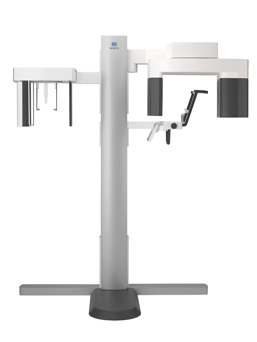
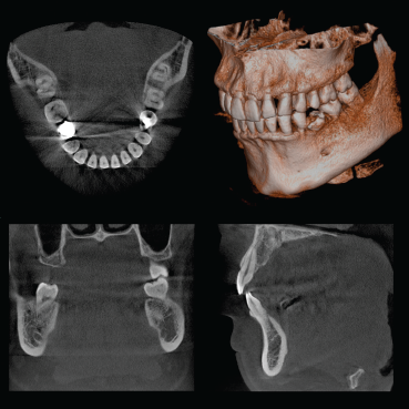
3D Accuitomo 170
From superior image quality to years of use, the Accuitomo features and sustainability are no comparison.

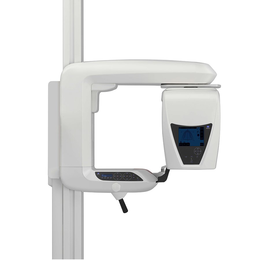
Veraview X800
Multifunctional, perfect for small spaces. Great quality, easy to use and ready to install.
Veraviewepocs 3D R100
Multifunctional unit great for the general dentist or specialist with six 3D fields of view.
3D Accuitomo 170
CBCT by Morita
Morita’s Most Advanced CBCT Unit
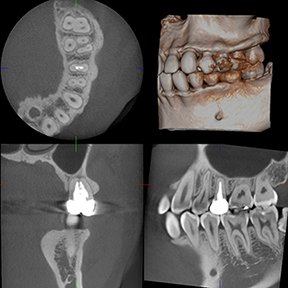
The 3D Accuitomo 170 CBCT is Morita’s most advanced unit. Highly refined, it is the 4th generation of the Accuitomo product line. It offers a voxel size of just 80 µm and displays even the most subtle details of bone structure. This super-fine voxel combined with the unit’s 14 bit grayscale capability creates a wide dynamic range which produces the highest quality visualization of both hard and soft tissue areas.
-
With the flat panel detector, high-quality and detailed Images of the structures of the head and neck can be generated. Adjusting the position of the FPD reduces X-ray dose, provides higher resolution, and minimizes distortion.
Optimizing collimation of the beam, depending on the size of the area, also reduces X-ray dose and X-ray scattering.
-
There are nine sizes for exposure regions with diameters ranging from 40 mm to 170 mm.
Ø 170 x 120 mm
Ø 170 x 50 mm
Ø 140 x 100 mm
Ø 140 x 50 mm
Ø 100 x 100 mm
Ø 100 x 50 mm
Ø 80 x 80 mm
Ø 60 x 60 mm
Ø 40 x 40 mm
Resolution stays high and distortion is minimized for all regions from the smallest (ø 40 x 40 mm) to the largest (ø 170 x 120 mm).
-
High-Resolution Mode (Hi-Res)
This is the highest resolution. Exposures are made at one-fourth the size of the detector pixels for the greatest spatial resolution. Ideal for observation of delicate bone structures such as the ossicular chain.
High-Fidelity Mode (Hi-Fi) This mode has high data density data to make clearer and sharper images. This is especially good for performing zoom reconstructions.
High-Speed Mode (Hi-Speed) Full scan: 10.5 sec. Half scan: 5.4 sec. Reduces motion artifacts. Good for children or others with difficulty remaining motionless.
Standard Mode (Std)
Suitable for limited and wide views of temporal bone, paranasal sinus, maxilla and mandible, individual teeth, etc.
-
The scout positioning system is easy and accurate. Use the triple beam positioning system for even greater precision.
Two-Direction Scout The region of interest can easily be targeted by making images from two directions. Then you can simply click on the images to specify the center of the region of interest. This information is transmitted to the X-ray unit, and the chair automatically moves into position.
Use scout to accurately determine the minimal region of interest before exposing the patient to the higher dosage CBCT scan.
Easy High Precision The region of interest can easily be targeted using the three positioning laser beams. The patient’s head is safely and securely stabilized by the chinrest and headrest.
-
Installing i-Dixel software on all intra-clinic computers enables sharing of image data on each linked client computer. Observation of images on non-network computers can be achieved with the One Data Viewer, and the One Volume Viewer without installing i-Dixel.
One Data Viewer & One Volume Viewer Software These unique Morita applications let you view three dimensional images and volume rendered images even if the computer does not have i-Dixel software installed.
CBCT data can be exported from the i-Dixel application and later stored on a DVD. This DVD can then be used on a computer outside the clinic to view CBCT images, volume rendered images and patient information.
Additional functions include zoom, black and white reverse, brightness, and contrast adjustment as well as optional length and angle measurement capabilities.
i-Dixel conforms to the following DICOM standards: 1.Modality worklist management service class (optional)
2. Storage service class
3. Modality performed procedure step service class
4. Print management service class

Veraview X800 CBCT/Pan/Ceph by Morita
The New Frontier for CBCT Imaging
The Veraview X800 is a multifunctional CBCT X-ray unit that produces stunning images for evaluation. High resolution, this unit offers a minute voxel size of just 80 μm and features an adjustable horizontal X-ray beam for artifact reduction. Two exposure modes offer control and flexibility with a 360° high-definition scan, or a faster 180° rotation with reduced dose. The X800 has reached the pinnacle of CBCT imaging technology.
-
For FOV Ø 40 × H 40 exposures, the voxel size is 80 μm and resolution is 2.5 LP/mm.
Spatial resolution indicates how small objects can be and still be discriminated visually. This is called spatial frequency and is usually expressed as “line pairs per millimeter (LP/mm)”. This indicates how many pairs of white and black lines can be discriminated within one millimeter; the higher the number, the greater the resolution.
MTF (Modulation Transfer Function) is one way to objectively evaluate the line-pair resolution and objectively express how many line-pairs and at what level of contrast can be discriminated. Generally, if MTF is 10%, naked eye discrimination is possible. Spatial resolution does not depend only on voxel size.
-
For the first time, Morita’s Zoom Reconstruction feature is available on a multi-functional unit. After taking an image with a voxel size of 125 μm, reconstruction can be repeated for a higher resolution of 80 μm voxel size without retaking the exposure.
-
A new taller field of view makes DICOM registration with surgical navigation systems even easier and more accurate.
-
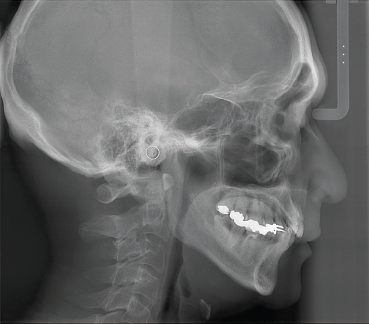
The Veraview X800 delivers exceptional panoramic and cephalometric imaging with precision and flexibility. Its adjustable flat panel detector optimizes X-ray beam alignment—an exclusive MORITA feature—ensuring ideal exposure for both panoramic and 3D scans.
Panoramic imaging offers two modes: a fast 7.4-second scan or a 14.8-second high-definition option, enhanced by features like Adaptive Focal Point/Grayscale, and Auto Image Enhancement for consistently sharp, detailed results.
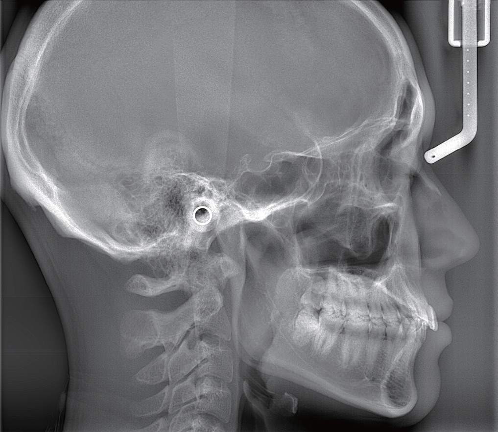
For cephalometric imaging, the unit provides both posterior-anterior and lateral views with a rapid 3.5-second scan time, plus a partial scan option for reduced radiation exposure.
-
Laser beam positioning is more accurate if you have good communication with the patient.
-
The control panel moves freely so that it can be used from the front or side, for improved access during patient positioning.
-
The chinrest can be lowered to approximately 34″ (865 mm) (short column) to accommodate patients in wheelchairs.


3D Imaging For Any Practice
(X700) The Veraviewepocs 3D R100 delivers outstanding 3D image quality with a built-in Dose Reduction Mode to enhance patient safety. It produces sharp, high-resolution images (125 μm voxel), clearly revealing bone structure, resorption, apical lesions, root fractures, and more—all with a fast 9.4-second scan time.
Multifunctional, it also offers panoramic and cephalometric imaging. This unit is a great solution for general dentistry and specialties.
Veraviewepocs 3D R100 CBCT/Pan/Ceph by Morita
-
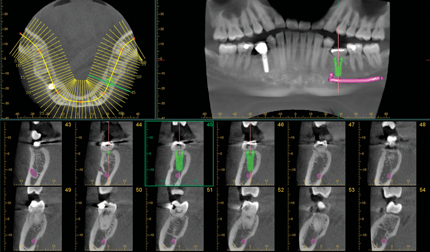
A total of six exposure areas from 40 x 40 mm up to 100 x 80 mm are available for various diagnostic needs. The full arch scan captures the maxilla and/or the mandible, and two height options of 50 or 80 mm.
Ø 100 x H 80 mm (3D Reuleaux Full Arch FOV)
Ø 100 x H 50 mm (3D Reuleaux Full Arch FOV)
Ø 80 x H 80 mm
Ø 80 x H 50 mm
Ø 40 x H 80 mm
Ø 40 x H 40 mm -
With a panoramic Scout, two-direction Scout, and five positioning laser beams—the R100 offers you multiple options for easy, reliable positioning and selection of the region of interest for 3D images.
-
Dose Reduction Mode automatically lowers radiation in areas of lower bone density, reducing exposure by up to 40% while improving soft tissue visibility, particularly in the anterior region.
-
The R100 offers many innovative features for brilliant image quality and effective dose reduction. Autofocus positioning ensures consistent, precise images with minimal effort, while real-time exposure adjustment improves contrast and dynamic range. Other features include panoramic focal plane adjustment and partial pan/ceph image functions.
Cephalometric programs include posterior-anterior and lateral. This unit’s high-speed performance requires only 2.6 to 5.8 seconds for a lateral projection.
Lasers
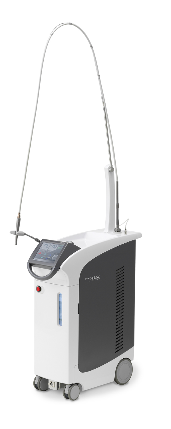
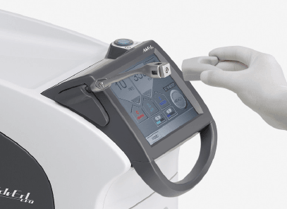
AdvErL EVO, Er:YAG Laser by Morita
Refined, Practical, and Clinically Effective
Morita’s AdvErL EVO, an Er:YAG laser, is the result of 30 years of research and development. Operating at a wavelength of 2,940 nm, Er:YAG vaporization is concentrated at the surface of the tissue, so the energy does not penetrate and damage deeper layers. It offers a sleek, modern design with built-in air and water systems for easy installation.
-
AdvErL EVO operates at a wavelength highly absorbed by water, making it ideal for precise, minimally invasive procedures. It excels in both hard and soft tissue applications with benefits such as reduced vibration, bacterial reduction, and faster healing without significant thermal damage.
Removal of subgingival calculi
Soft tissue curettage
Gingival incision and excision
Hemostasis and coagulation
Frenectomy and frenotomy
Ablation, vaporization
Class I, II, III, IV and V cavity preparation
Caries removal
-
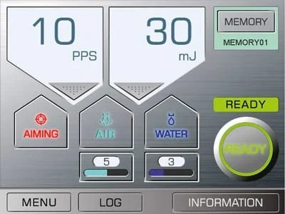
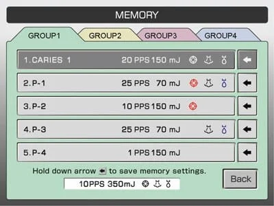
The display is large, easy to read, and has an intuitive design. All settings can be easily confirmed at a glance. There are 20 pre-programmed settings that can easily be retrieved. Additionally, a wide range of tip options is available.
-
Lasers can be a powerful adjunct in modern endodontic therapy—particularly in complex canal anatomies where conventional methods may fall short. AdvErL EVO applies the principle of laser-activated irrigation for effective treatment of the root canal anatomy.
Applications include:
Root canal access and preparation
Debridement and cleaning
Assisting in achieving patency
-
AdvErL EVO is ideal for periodontology. Most notably, this unit has been studied and recognized clinically for the effective treatment of peri-implantitis, a challenging disease leading to bone loss around an implant. Applications include:
Implant recovery
Regeneration of tissue and permanent removal of bacteria with a low-heat treatment process
Soft tissue curettage
Sulcular debridement and removal of subgingival calculus
Removal of inflamed or edematous tissue and granulation tissue from bony defects
What people are saying
“Let me start by saying that the ease-of-use and excellent quality of this device. Not only is the CT scanner incredibly user-friendly, but it also produces highly accurate CT scans. The level of detail it captures is truly impressive.”